Hypodense (less dense): If an abnormality is less dense than the reference structure, we would describe it as hypodense.
What is a small Hypodensity?
Abstract. Small hypodense renal lesions with a round shape are frequently detected on CT scans of the upper abdomen after contrast medium administration. In nearly all cases these round hypodensities are simple small cysts with no clinical significance.
Is Hypodensity a lesion?
Hypodense splenic lesions are frequently encountered on abdominal CT images. Although most hypodense lesions of the spleen can be considered benign, some findings and clinical conditions warrant closer attention to the lesion.
What is Hypodensity of white matter?
We conclude that hypodensity of the cerebral white matter in patients with transient ischemic attack or minor stroke is associated with an extra risk of future stroke, from large as well as from small vessels, and particularly in patients under 70 years old; this increase of risk is independent of other risk factors
What is hyperdense on CT?
Hyperdensity at CT was due to the high hemoglobin content of retracted clot or sedimented blood. The various patterns seen can be related to sequential changes occurring in blood following hemorrhage. Relative hyperdensity and its variations seen on precontrast scans are useful diagnostic signs of recent hemorrhage.
Are hypodense lesions cancerous?
concluded that finding a small, hypodense lesion in the liver in a patient with otherwise no definite metastases was a benign finding. Krakora et al. [21], in a study of 153 patients with breast cancer, discovered small hypoattenuating hepatic lesions in 35%.
Are cysts Hypodense?
The central cystic component appears hypodense on unenhanced CT and on all phases of contrast-enhanced dynamic examination (Fig. 8). The presence of a fluid–fluid level within a cavernous hemangioma is a usual variant that has been described in the literature.
How do you read a CT brain?
Examine the brain for:
Symmetry – make sure sulci and gyri appear the same on both sides. Grey-white differentiation – the earliest sign of a CVA on CT scan is the loss of the grey-white interface on CT scan. Shift – the falx should be in the midline with ventricles the same on both sides.
What is hyperintense signal?
A hyperintensity or T2 hyperintensity is an area of high intensity on types of magnetic resonance imaging (MRI) scans of the brain of a human or of another mammal that reflect lesions produced largely by demyelination and axonal loss.
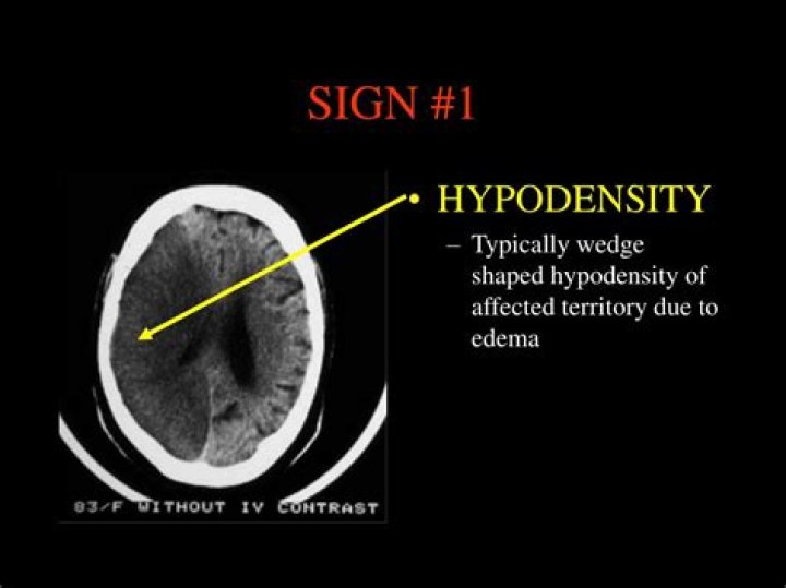
What does Hypodensity in brain mean?
Objective: Hypodense lesions identified on computed tomographic (CT) scans are often assumed to indicate ischemia. The purpose of this study was to investigate regional cerebral blood flow (rCBF) in hypodense areas of the brain after severe traumatic brain injury.
Should I be worried about liver lesions?
What are liver lesions? Liver lesions are abnormal growths that may be noncancerous (benign) or cancerous. Benign lesions occur for a variety of reasons and are typically not cause for concern.
What does low attenuation on a CT scan mean?
BACKGROUND The low attenuation areas on computed tomographic (CT) scans have been reported to represent emphysematous changes of the lung. However, the regional distribution of emphysema between the inner and outer segments of the lung has not been adequately studied.
What are the symptoms of white matter disease?
Symptoms of white matter disease may include:
issues with balance.walking slow.more frequent falls.unable to do more than one thing at a time, like talking while walking.depression.unusual mood changes.
Is mild white matter disease serious?
This loss may be the result of an injury, infection, or underlying health condition. Mild cases of brain atrophy may have little effect on daily functioning. However, brain atrophy can sometimes lead to symptoms such as seizures, aphasia, and dementia. Severe damage can be life threatening.
Can white matter lesions in the brain be nothing?
White matter lesions observed on brain MRI are usually characteristic and occur in specific areas including the corpus callosum and pons. “However, in many cases, the white matter lesions as isolated observations are nonspecific” and could be due to MS or another cause, explained Drs Lange and Melisaratos.
What does Hyperdense mean?
Hyperdense definition
(medicine) Extremely dense. A hyperdense liver. adjective.
What does Hyperdense cyst mean?
Conclusions: Hyperdense renal cyst is a simple cyst which has suffered bleeding or infection. Diagnosis is made by CT scan and it does not require treatment.
Why is blood white on CT scan?
Acute haemorrhage absorbs X-rays and appears hyperdense (white) on CT scans. As the clot retracts it becomes more hyperdense over the first few hours up to 7 days; then isodense with brain over the following 1-4 weeks and finally hypodense compared with brain over the subsequent 4-6 weeks.